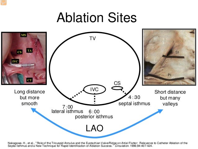


In panels F and G, red line indicates CTI ablation line, green circle indicates possible haemorrhage, and yellow circle indicates possible damage due to positioning of the catheter in the CS. In conclusion, this case demonstrates that iCMR-guided catheter ablation is feasible and allows for real-time anatomic visualization and advanced characterization of ablation effects.Īrrows indicate regions of interest. Lastly, 3D reconstructions derived from the 3D LGE-acquisition were made of the RA ( Panel G) and LA ( Panel H) to visualize the ablation lesions. Also, an enhanced area near the ostium of the coronary sinus (CS) was observed (yellow dotted circle), which might indicate myocardial damage due to extensive CS-catheter manoeuvering. This might indicate an ablation core with acute myocardial haemorrhage. An area of low signal intensity is visible in the middle of the CTI ablation line ( Panel F, green dotted circle). Post-ablation 3D late gadolinium enhancement (LGE) images show both acute right atrial (RA) CTI ablation lesion ( Panel C) as well as chronic left atrial (LA) pulmonary vein lesions resulting from the previous cryo-balloon ablation ( Panel D). T2-weighted images show an increased wall thickness and enhanced signal intensity indicating extensive oedema at the CTI region compared with pre-ablation ( Panels A and B). Immediately after ablation, conventional CMR imaging was performed to visualize and characterize the acute ablation lesion. However, due to recent radiation exposure, the patient was proposed for flutter ablation in a magnetic resonance imaging (MRI) environment as an alternative to X-ray fluoroscopy.Ī complete isthmus block was achieved after an uncomplicated MRI-guided radiofrequency CTI ablation.

In August, the patient presented with symptomatic atrial flutter and was referred for CTI ablation. Prior cardiac history included atrial fibrillation, for which he underwent cryo-balloon pulmonary vein isolation in June 2022. The present case demonstrates a real-time interventional cardiac magnetic resonance imaging (iCMR)-guided cavotricuspid isthmus (CTI) ablation performed in a 53-year-old male patient.


 0 kommentar(er)
